
What Histopathologists Do
Medical students and foundation doctors often have limited exposure to histopathology, which is why there are a lot of misconceptions about what a histopathologist actually does.
Here is a brief overview. If it whets your appetite, contact your nearest Pathology Ambassador or your local histopathology department and arrange to meet with one of the trainees/consultants who will be happy to answer your questions and show you around.
Histopathologists have a hugely varied and diverse workload. Within a single day, a histopathologist may do any/all of the following:
'Cut up/grossing' (i.e. when surgical specimens are received)
The pathologist:
- Describes the specimen’s macroscopic features, e.g. size, weight, perforations, adhesions etc.
- Chooses whether to ink the specimen. The coloured ink can be seen when the tissue is viewed microscopically, allowing orientation (e.g. green denotes anterior surface) or demarcating an external surface.
- Slices the specimen, examines the slices and describes key features, e.g. in the case of a tumour: size, location within the specimen, relationship to the resection margins, whether it macroscopically invades blood vessels/lymph nodes etc.
- Chooses which slices should be processed and made into formalin fixed paraffin embedded (FFPE) tissue blocks (as large specimens cannot be examined in their entirety). Accurate microscopic diagnosis relies on selection of the most appropriate blocks.
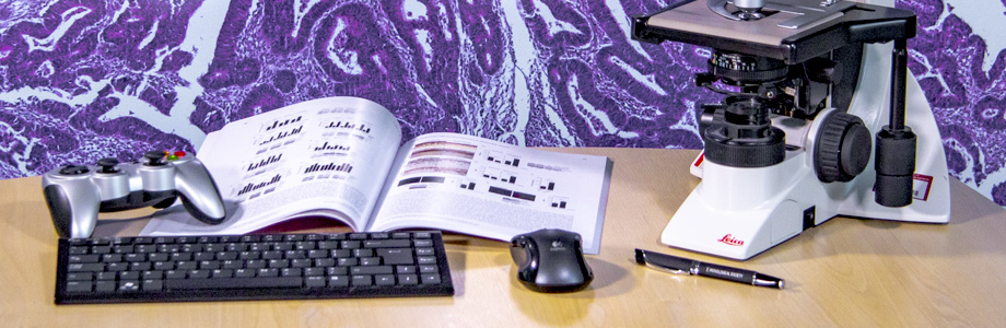
Microscopy
Thin sections of FFPE tissue are placed onto glass slides and stained so that the tissue can be visualised microscopically (either directly using a microscope, or the slides may be scanned and viewed on a computer). Tissue may come from resections, biopsies, or cell suspensions (cytology).
The pathologist:
- Decides the diagnosis.
- Records features which may have therapeutic/prognostic implications (e.g. tumour type, grade, lymph node involvement, resection margin involvement etc.)
- May request special stains (e.g. to diagnose pathogens) or molecular tests (e.g. to direct treatment, determine genetic risk etc.)
Report writing & the MDT
The pathologist:
- Decides the diagnosis.
- Writes a histopathology report, recording the relevant macroscopic and microscopic features and incorporating information from the clinical history and medical notes.
- Presents the report at the multidisciplinary team meeting (MDT). Here information from the pathologist, surgeons, radiologists and oncologists is synthesised to determine the optimum management plan for the patient.
Frozen Sections
A frozen section may be requested when pathology information is required urgently in order to make an intra-operative decision (e.g. type of lesion during resection of a probable tumour). The pathologist examines the tissue macroscopically and selects which part to process; this is frozen (so that it is firm, even in the absence of FFPE which is too time-consuming) and sectioned (thin sections are cut, placed onto glass slides and stained). The pathologist examines the tissue microscopically and contacts the surgical team urgently with the information they require.
Post mortems
In the first two years of histopathology training, trainees are taught how to perform a post mortem and must perform a minimum number of post mortems. Subsequently, post mortems are optional with an optional autopsy exam.
The pathologist:
- Conducts an external examination e.g. looking for signs of disease, injury, markers of identification etc.
- Removes the internal organs, weighs and examines them, recording any pathological features.
- May take tissue for microscopic examination.
- Decides the cause of death. Writes the death certificate and post mortem report.
- May be asked to present the findings at a Coroner’s Court.
Other Activities
- Teaching of medical students, histopathology trainees, biomedical scientists etc.
- Leadership/management roles: within the hospital, Deanery, National Organisations e.g. the Royal College of Pathologists, the Pathological Society of Great Britain & Ireland etc.
- Research
- Audit
- Public/patient engagement