
The future of histopathology
Histopathology is not a static specialty – exciting developments are currently underway:
- Virtual/digital autopsies: These are being carried out in certain centres. A pathologist performs an external examination and the body is then imaged e.g. in a CT scanner. If a cause of death is identified by imaging, evisceration (removal of the internal organs) is avoided.
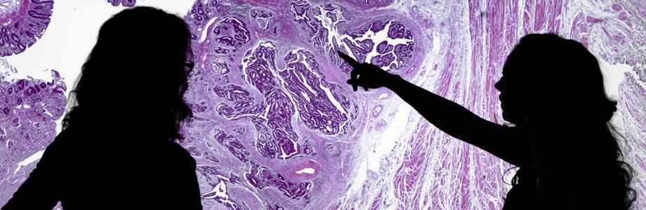
- Digital pathology: This is being introduced in certain areas. Glass histology slides are scanned and the images are viewed by a pathologist using a computer rather than a microscope. This enables slides to be viewed remotely, anywhere in the world, and archived digitally (for examples, see http://www.virtualpathology.leeds.ac.uk/slides/library/ ).
- Molecular testing: More and more molecular tests are becoming available and integrated into the pathology report. These tests can have implications for treatment, prognosis or reveal familial genetic risk for certain diseases.
- Change in working: In some centres scientists undertake cut-up and selective histology reporting.